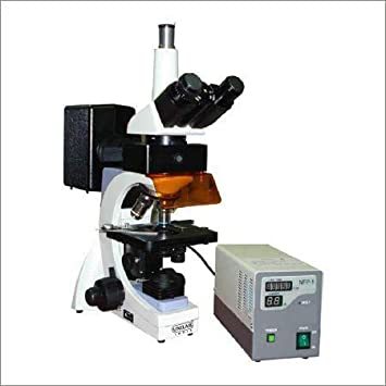
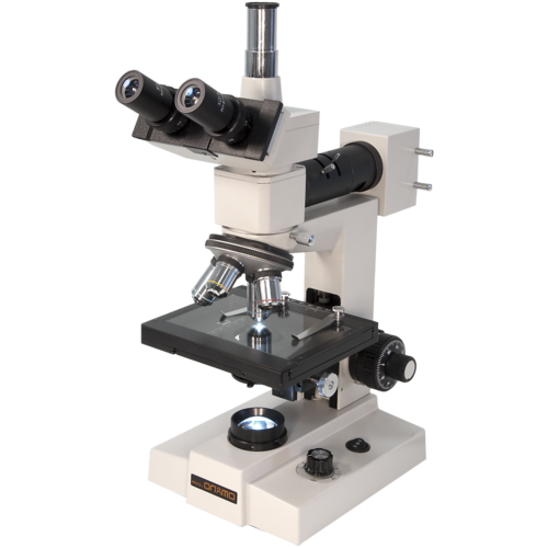
About Trinocular Fluorescence Microscope
Specifications same as model BTM 423 butprovided with Trinocular Head with 45 deg. inclined binocular observation andstraight tube for Photomicrography
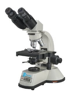
A Robust and stable die-caststable body with Co-Axial coarse and fine focusing mechanism based on four step reeducation gears system which runs on ball- bearing guide ways with tension control ring.Stopper system is provided so that the slides may not damage.
Binocular Head Inclined at 45/30deg.With all Anti-Fungal Coating prisms. Ratable 360 deg. IPD adjustment between54mm -74mm.
Nose Piece A high precision quadruple nose piece running on Balls with positive indexing.
All positions are parcentered and personalized
Mechanical Stage- Low positioned Co-axial calibrated double plate mechanical with feather touch movement. Wide Field eye piece.
Epi Fluorescent System: The Epi-Illuminationis through 100W Ultra High pressure Mercury Lamp in a Lamp House attached to the Fluorescence Filter Block.
Filters -Two Exciting filters B (Blue), G (Green) Exciting light Filter system, O Ordinary Light System.
Protection Barrier- Orange coloured plastic screen protection barrier to resist the Ultra Violet Light.
Eye Piece- High Eye Point Anti Fungal Extra Wide Field 10x /FN 18mm) paired eye piece
Fluorescence Objective: 4x,10x,40x(SL) ,100xOil(SL) with Immersion Oil Fluorescence Free Power : AC input 220V,Indicator Display.
Crystal Clear Imaging and Versatile MagnificationWith a magnification range of 40x to 1000x, and a maximum digital camera resolution of 1920 x 1080 pixels, this trinocular fluorescence microscope offers remarkable clarity for detailed observations. Achromatic objectives and adjustable wide field eyepieces minimize aberration and enhance image fidelity, while its three viewing ports allow simultaneous visual and digital monitoring.
Flexible Camera Integration and Live ViewingDesigned with a dedicated trinocular port, this microscope is fully compatible with CMOS cameras, permitting real-time video streaming and high-resolution still image capture (up to 12MP, depending on the camera). JPEG and PNG file formats are supported, and users can enjoy a live view at a smooth 30 fps for dynamic sample monitoring.
Advanced Illumination for Reliable ResultsChoose between a powerful 100W mercury vapor lamp or energy-efficient 3W LED light to accommodate a range of fluorescence techniques. The system supports both transmitted and incident light, ensuring samples are brightly and evenly illuminated for optimal visualization during fluorescence applications.
FAQs of Trinocular Fluorescence Microscope:
Q: How do I attach a digital camera to the trinocular fluorescence microscope?
A: A digital camera connects via the dedicated trinocular port, which is designed to be compatible with most CMOS cameras. Simply mount your camera on the port and secure it for both live view and image capture functionalities.
Q: What benefits does the microscopes trinocular head provide?
A: The trinocular head allows for simultaneous visual observation and digital imaging, enabling documentation, analysis, and sharing of results without disturbing the ongoing observation or experiment.
Q: When should I use the mercury vapor lamp versus the LED illumination?
A: Use the 100W mercury vapor lamp for applications demanding intense fluorescence excitation across multiple wavelengths. The energy-efficient 3W LED is recommended for less demanding fluorescence or when heat-sensitive samples are being examined.
Q: Where can I obtain spare parts or replacement components for this microscope?
A: Spare bulbs, fuses, objectives, and stage clips are available through the manufacturer, exporter, or authorized suppliers. Always consult your product documentation or supplier for recommended sources.
Q: What is the maximum resolution for imaging and video capture with camera attachment?
A: Still images can be captured at up to 12MP, depending on the camera, while video capture supports full HD 1080p resolution at 30 frames per second, suitable for both documentation and presentations.
Q: How do I adjust the focus for precise sample observation?
A: The microscope is equipped with a coaxial coarse and fine focusing system, offering a 25 mm coarse range and 0.002 mm fine adjustment per division, along with tension adjustment, enabling meticulous focusing for high-precision work.
Q: What are the main steps in setting up the microscope for fluorescence observation?
A: Mount the sample on the double-layer stage, select the appropriate objective lens, adjust illumination with the preferred light source, and use the fine focus to bring the sample into sharp view. Attach a camera if digital imaging or live monitoring is needed.


 English
 Spanish
 French
 German
 Italian
 Chinese (Simplified)
 Japanese
 Korean
 Arabic
 Portuguese
 Send Inquiry
 Get Latest Price
